It works closely with a globular protein called actin that polymerizes to create actin filaments. Myosin is a special protein that converts adenosine triphosphate (ATP), a molecule that cells use in order to live and work, into mechanical energy (energy for work).Also question is, what is the function of myosin in muscle contraction?
Myosin Molecules and Thick Filaments Myosin is a motor protein that generates the force in a muscle contraction much like the stroke of an oar. It consists of a head and a tail region. Together, the tails of approximately three hundred myosin molecules form the shaft of the thick filament.
Similarly, what Proteins make up myosin? Thick filaments contain myosin, thin filaments contain actin , troponin and tropomyosin. Scientists think that muscles contract by the two types of filament sliding over each other so that they overlap more (Figure 5). The sturcture of myosin in thick muscle filaments. Myosin is made up of six polypeptide chains.
Accordingly, where is myosin protein found?
Myosin is a superfamily of proteins which bind actin, hydrolyze ATP and transduce force. Thus most are located in muscle cells. Composed of head, neck and tail domains. Head domain binds the actin and moves along it.
How does myosin work as a motor protein?
Myosins are a superfamily of actin motor proteins that convert chemical energy in the form of ATP to mechanical energy, thus generating force and movement. The myosin heads bind and hydrolyze ATP, which provides the energy to walk toward the plus end of an actin filament.
Why is myosin so important?
Myosins are often referred to as molecular motors because they use energy to move. Myosin proteins are involved in many cellular functions. Their ability to transport materials and create force through contractions make them important in the process of cell division. Myosins are also involved in cell movement.What is myosin made of?
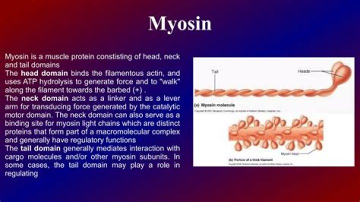
Structure and functions Most myosin molecules are composed of a head, neck, and tail domain. The head domain binds the filamentous actin, and uses ATP hydrolysis to generate force and to "walk" along the filament towards the barbed (+) end (with the exception of myosin VI, which moves towards the pointed (-) end).What are the 6 steps of muscle contraction?
Help me put the 6 steps of muscle contraction in order? - Ca2+ is pumped back into the terminal cisternae. C)
- Myosin heads bind to the binding sites on the actin. D)
- ATP is hydrolyzed and re-energizes the myosin head. E)
- ATP causes the myosin head to be released by binding to the myosin head.
- Ca2+ is released from the terminal cisternae (end of motor neuron)
How do you explain muscle contraction?
Muscle contraction is the activation of tension-generating sites within muscle fibers. In physiology, muscle contraction does not necessarily mean muscle shortening because muscle tension can be produced without changes in muscle length such as holding a heavy book or a dumbbell at the same position.What is the correct order of steps in muscle contraction?
The process of muscular contraction occurs over a number of key steps, including: - Depolarisation and calcium ion release.
- Actin and myosin cross-bridge formation.
- Sliding mechanism of actin and myosin filaments.
- Sarcomere shortening (muscle contraction)
What is the role of ATP in muscle contraction?
What is the role of ATP in muscle contraction? ATP is responsible for cocking (pulling back) the myosin head, ready for another cycle. When it binds to the myosin head, it causes the cross bridge between actin and myosin to detach. ATP then provides the energy to pull the myosin back, by hydrolysing to ADP + Pi.What are the major muscles of the human body?
The major skeletal muscle groups forming the upper body are the abdominal, pectoral, deltoid, trapezius, latissimus dorsi, erector spinae, biceps, and triceps. The major skeletal muscle groups of the lower body are the quadriceps, hamstrings, gastrocnemius, soleus, and gluteus. Muscles move by contracting.How are muscles activated?
In experiments, muscles are typically activated by electric stimuli applied to muscle surface or to the nerve innervating the muscle. If the strength of a single stimulus exceeds a certain threshold, the muscle responds by a brief period of contraction followed by relaxation (twitch).Is DNA a protein?
Today, proteins are formed following instructions given by DNA (deoxyribonucleic acid) which in turn is synthesized by specific enzymes that are proteins. DNA contains the genetic information of all living organisms. Proteins are large molecules made up by 20 small molecules called amino acids.How many types of myosin are there?
Myosin V exists in three isoforms – Va, Vb, and Vc – but only the first two are highly expressed in nervous tissue. All isoforms are dimers with a long neck region that binds multiple light chains (mainly calmodulin) and a globular tail portion that contributes to cargo binding (Figure 2).Is actin a protein?
Actin is a family of globular multi-functional proteins that form microfilaments. An actin protein is the monomeric subunit of two types of filaments in cells: microfilaments, one of the three major components of the cytoskeleton, and thin filaments, part of the contractile apparatus in muscle cells.What is the largest protein?
Titin
Who discovered myosin?
The Beginning. A viscous protein was extracted from muscle with concentrated salt solution by Kühne (1864), who called it “myosin” and considered it responsible for the rigor state of muscle.What are five proteins found in muscle?
The most important are the contractile proteins actin and myosin. Among the regulatory proteins, troponin, tropomyosin, M-protein, beta-actin, gamma-actin and C-protein are great importance.What are the different types of motor proteins?
Just three families of motor proteins—myosin, kinesin, and dynein—power most eukaryotic cellular movements (Fig. 36.1 and Table 36.1). During evolution, myosin, kinesin, and Ras family guanosine triphosphatases (GTPases) appear to have shared a common ancestor (Fig.How are Microfilaments formed?
Microfilaments are formed when globular (g)-actin-monomers polymerize into filamentous (f) actin polymers. Rapid addition of monomers at the membrane end is the process used in the formation of pseudopodia for cell migration. The rate of polymerization is regulated by calcium, ATP, camp, and actin binding proteins.What is keratin protein?
Keratin is the type of protein that makes up your hair, skin, and nails. Keratin is a protective protein, less prone to scratching or tearing than other types of cells your body produces. Keratin can be derived from the feathers, horns, and wool of different animals and used as an ingredient in hair cosmetics.