Then, what is the function of the pharyngeal constrictor muscles?
Function. As soon as the bolus of food is received in the pharynx, the elevator muscles relax, the pharynx descends, and the constrictors contract upon the bolus, and convey it downward into the esophagus. They also have respiratory mechanical effects.
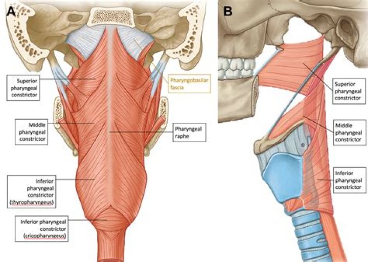
Furthermore, what passes between superior and middle pharyngeal constrictors? Between the superior and middle pharyngeal constrictor muscles, the stylopharyngeus muscle, the glossopharyngeal nerve and the stylohyoid muscle pass through. Between the middle and inferior pharyngeal constrictor muscles, the internal laryngeal nerve and the superior laryngeal artery and vein pass through.
Secondly, where is the superior pharyngeal constrictor muscle located?
The superior constrictor muscle is located anterior to the prevertebral muscles and posterior to the buccinator muscle, from which it is separated by the pterygomandibular raphe.
What does the Salpingopharyngeus do?
As the salpingopharyngeus is used to open the eustachian tubes to equalize pressure in the middle ear, the muscle can easily be stimulated by swallowing. The salpingopharyngeus is innervated by the vagus nerve via the pharyngeal plexus, and irrigated by the ascending pharyngeal artery.
What pharyngeal muscles are involved in swallowing?
The pharynx is pulled upwards and forwards by the suprahyoid and longitudinal pharyngeal muscles – stylopharyngeus (IX), salpingopharyngeus (pharyngeal plexus—IX, X) and palatopharyngeus (pharyngeal plexus—IX, X) to receive the bolus.Where is the larynx located?
Larynx: A tube-shaped organ in the neck that contains the vocal cords. The larynx is about 5 cm (2 in.) long. It is part of the respiratory system and is located between the pharynx and the trachea.What does the Stylopharyngeus do?
The stylopharyngeus: elevates the larynx. elevates the pharynx. dilates the pharynx to permit the passage of a large food bolus, thereby facilitating swallowing.What is the pharyngeal?
The pharynx (plural: pharynges) is the part of the throat behind the mouth and nasal cavity, and above the esophagus and larynx – the tubes going down to the stomach and the lungs. It is found in vertebrates and invertebrates, though its structure varies across species.What are the pharyngeal muscles?
The pharyngeal muscles are a group of muscles that form the pharynx, which is posterior to the oral cavity, determining the shape of its lumen, and affecting its sound properties as the primary resonating cavity. The pharyngeal muscles(involuntary skeletal) pushing the food into the esophagus.What kind of muscle is the Cricopharyngeus?
inferior constrictorHow many muscles are in the throat?
Some 50 pairs of muscles and many nerves work to receive food into the mouth, prepare it, and move it from the mouth to the stomach. This happens in three stages. During the first stage, called the oral phase, the tongue collects the food or liquid, making it ready for swallowing.How many larynx do we have?
The laryngeal skeleton consists of six cartilages: three single (epiglottic, thyroid and cricoid) and three paired (arytenoid, corniculate, and cuneiform).What is in the retropharyngeal space?
The retropharyngeal space (between the posterior pharyngeal wall and the prevertebral layer of deep cervical fascia) contains loose connective tissue and lymph nodes that drain the nasopharynx, paranasal sinuses, middle ear, teeth, and adjacent bones.What organ is controlled by the Styloglossus muscle?
| Styloglossus | |
|---|---|
| Insertion | tip and sides of tongue |
| Artery | sublingual branch of the lingual artery. |
| Nerve | Hypoglossal nerve (CN XII) |
| Actions | retraction and elevation of tongue |
