Diaphragm anatomy and function The diaphragm is a thin skeletal muscle that sits at the base of the chest and separates the abdomen from the chest. When you exhale, the diaphragm relaxes and the air is pushed out of lungs. It also has some nonrespiratory functions as well.Similarly one may ask, are respiratory muscles skeletal muscles?
Skeletal Muscle Physiology. The pump muscles involved in respiration are skeletal muscles. Because of these functional demands, the diaphragm muscle is much more active than other skeletal muscles (e.g., limb muscles not involved in posture).
Similarly, what does diaphragm pain feel like? Symptoms of diaphragm pain a “stitch” in your side when you exercise. inability to take a full breath. low blood oxygen levels. pain in your chest or lower ribs.
Likewise, people ask, what are the muscles of the diaphragm?
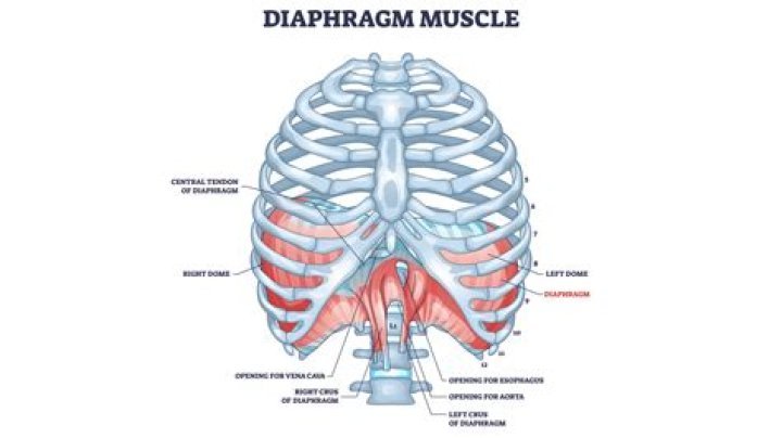
The diaphragm is a musculotendinous sheet. It has three muscular parts (sternal, costal, and lumbar), each have their own origin and all insert into the central tendon of diaphragm. The diaphragm is shaped as two domes, with the right dome positioned slightly higher than the left because of the liver.
Can the diaphragm cause pain?
It is the primary muscle that the body uses when breathing. The diaphragm moves downward so the lungs can fill with air during inhalation. People may sometimes feel pain or discomfort in their diaphragm, although in some cases it is possible that the pain is coming from a different, nearby body part.
Do lungs have muscle?
The lungs have no skeletal muscles of their own. The work of breathing is done by the diaphragm, the muscles between the ribs (intercostal muscles), the muscles in the neck, and the abdominal muscles. As the diaphragm contracts, it increases the length and diameter of the chest cavity and thus expands the lungs.What muscles are used in expiration?
During active expiration, the most important muscles are those of the abdominal wall (including the rectus abdominus, internal and external obliques, and transversus abdominus), which drive intra-abdominal pressure up when they contract, and thus push up the diaphragm, raising pleural pressure, which raises alveolarWhat muscles never stop working?
During expiration, the lungs deflate without much effort from our muscles. However, the expiratory muscles – internal intercostals, rectus abdominis, external and internal obliques, transversus abdominis – can contract to force air out of the lungs during active breathing periods.What muscles affect breathing?
The diaphragm is the major muscle responsible for breathing. It is a thin, dome-shaped muscle that separates the abdominal cavity from the thoracic cavity. During inhalation, the diaphragm contracts, so that its center moves caudally (downward) and its edges move cranially (upward).Can muscles collapse?
Causes of muscle atrophy. Unused muscles can waste away if you're not active. But even after it begins, this type of atrophy can often be reversed with exercise and improved nutrition. Muscle atrophy can also happen if you're bedridden or unable to move certain body parts due to a medical condition.Why do we use accessory muscles to breathe?
Accessory expiratory muscles are the abdominal respiratory muscles (rectus abdominis, transverse abdominis, and external and internal obliques). They augment the passive recoil of the lungs during expiration and also help in inspiration.What is respiratory muscle fatigue?
DEFINITION AND TYPES OF RESPIRATORY MUSCLE FATIGUE Muscle fatigue can be defined as a condition in which there is a reduction in the force generating capacity of the muscle resulting from muscle activity under load which is reversible by rest.What causes respiratory muscle weakness?
Some examples of conditions that cause muscle weakness are: ALS (amyotrophic lateral sclerosis, also known as Lou Gehrig's Disease), muscular dystrophy and conditions from genetic abnormalities. How can neuromuscular weakness affect my breathing? Many muscles are needed for normal breathing.What is the structure of the diaphragm?
The diaphragm is a C-shaped structure of muscle and fibrous tissue that separates the thoracic cavity from the abdomen. The dome curves upwards. The superior surface of the dome forms the floor of the thoracic cavity, and the inferior surface the roof of the abdominal cavity.Can the diaphragm be strengthened?
Diaphragmatic breathing is a type of a breathing exercise that helps strengthen your diaphragm, an important muscle that helps you breathe. This breathing exercise is also sometimes called belly breathing or abdominal breathing. It has a number of benefits that affect your entire body.What organ is under the diaphragm?
The liver is located under the ribs on the right hand side of the body. It lies just below the lungs, under the top of the diaphragm to which it is attached. The diaphragm is the muscle beneath the lungs which regulates our breathing. The liver is partly protected by the rib cage.Can you live without a diaphragm?
Kitaoka H(1), Chihara K. The diaphragm is the only organ which only and all mammals have and without which no mammals can live.What level is the diaphragm?
The diaphragm is located at the inferior-most aspect of the ribcage, filling the inferior thoracic aperture. It acts as the floor of the thoracic cavity and the roof of the abdominal cavity. The attachments of diaphragm can be divided into peripheral and central attachments.Can anxiety cause diaphragm spasms?
A diaphragmatic cramp or spasm can cause chest pain and shortness of breath that can be mistaken for a heart attack. Some people also experience sweating and anxiety during a diaphragm spasm. Vigorous exercise can cause the diaphragm to spasm, which often results in what people call a side stitch.How thick is the diaphragm?
Average thickness of the diaphragm in healthy volunteers is between 0.22–0.28 cm.What are the three openings in the diaphragm?
The diaphragm has three openings: Aortic Hiatus - the most dorsal opening, contains the aorta, azygous vein and thoracic duct. Oesophageal Hiatus - contains the oesophagus, dorsal and ventral vagal trunks. Caval Foramen - lies within the central tendinous region of the diaphragm and contains the caudal vena cava.Can you feel your diaphragm?
Place one hand on your upper chest and the other just below your rib cage. This will allow you to feel your diaphragm move as you breathe. Breathe in slowly through your nose so that your stomach moves out against your hand. The hand on your chest should remain as still as possible.